


Everyday Medicine: The Third Eyelid
Everyday Medicine: The Third Eyelid
Everyday Medicine is an intermittent series of blog posts highlighting tests, treatments and procedures commonly used at AMC. Some past examples of this type of blog post include “Vital Signs” and “The Highs and Lows of Blood Sugar.”
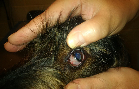
The other day I was examining a dog’s head. As part of the exam, I pushed on the dog’s upper eyelid to examine his third eyelid. When the white structure popped out of its hiding place under the lower lid, the owner gasped, and asked about the unexpected structure in her dog’s eye.
Nictitating Membrane
Another name for the third eyelid is nictitating membrane or haw. If a sick pet has its third eyelids exposed, veterinarians might say the pet has “haws.” The medical name comes from the Latin word nictare or blink. Most animals have a nictitating membrane, but primates have only a rudimentary one. Look in the mirror and the little curved slip of tissue in the inside corner of your eye is all you have left of your nictitating membrane.
Form
The nictitating membrane is translucent in birds, snakes and reptiles, but in dogs and cats, the membrane is opaque. Unlike the two outer eyelids, there is only one nictitating membrane and instead of moving up and down, the nictitans moves horizontally over the eye. The nictitans is part of the blink response in some animals, but not in dogs and cats, which is why I think my client was surprised about seeing something in her pet’s eye that she had not seen before. Most of the time, in a dog or cat, the nictitans is tucked behind the lower eyelid and not visible.
Function
The nictitans has important functions to protect the eye and thus vision. It removes debris from the eye. The nictitating membrane also provides protection for the eye when animals hunt, graze, feed their young, and encounter harsh conditions like snow and wind. Behind the nictitans is a tear gland that produces about half of the tears required to keep the eye moist. And, the nictitans helps to distribute the tears over the surface of the eye.
Disease
In general, the third eyelid is not prone to disease. “Cherry eye,” or protrusion of the tear gland normally found behind the nictitans, is the most common disorder of the third eyelid. Common in certain breeds of dogs and cats, the gland can easily be sutured back into place. Since the gland produces 50% of tears, its presence is critical to promoting a healthy eye. If you notice both third eyelids are up in your pet, this abnormality suggests a systemic illness. The nictitans may also protrude when your pet is recovering from anesthesia. Finally, because the face is frequently exposed to sunlight, the nictitans can rarely develop sunlight-induced squamous cell carcinoma. This tumor might appear as a reddening or lump protruding out from under the lower lid.
Amazing how important such a small and practically invisible organ can be to your pet’s healthy vision.
About the Author
Related Posts
-
Dogs EmergencyMarch 20, 2024

City Safety for Urban Dogs [2024 Update]
Learn More -
DogsMarch 13, 2024
The Impact of Dog Size, Breed, & Nose Length on Longevity
Learn More -
Dogs Internal MedicineDecember 06, 2023

The Surprising Link Between Respiratory and Gastrointestinal Diseases in Dogs
Learn More