CT Versus MRI: Battle of the Big Machines
CT Versus MRI: Battle of the Big Machines
Veterinarians at The Animal Medical Center depend on high tech equipment to make diagnoses and monitor treatment success. Two commonly used pieces of high tech equipment are the CAT scan or CT (Computed Tomography) and MRI (Magnetic Resonance Imaging). Often, if I recommend a CT, pet owners will ask if an MRI would be better. I checked with one of AMC’s board certified radiologists, Dr. Anthony Fischetti, to help dispel any myths about which test is the best. He says “both are equally as good, but which test is used depends on the body part being imaged and the type of resolution required to optimally image that body part.”
Big Machines at AMC
Computed tomography was introduced to human medicine in the 1970s. The AMC acquired its first CT scanner about 10 years later and is currently using its third scanner, a high-powered 64-slice CT scanner. Magnetic resonance imaging became commercially available in the 1980s and The AMC installed its first MRI machine in 2002 and upgraded that machine in 2006 for a more powerful model. To give you a comparison of the frequency of use of these tests, in 2007, a total of 73 million CT scans were performed on humans. In 2013, 700 CT scans and 600 MRI exams were performed – just at The AMC!
Starting at the Top
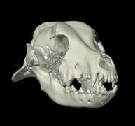
Imaging the head is a particularly good example of why we need both a CT scanner and an MRI machine at The AMC. The brain is composed of soft tissue and the boney skull is clearly hard tissue. When our neurologists want an image of the brain to determine the cause of seizures, they choose an MRI because it produces images with exquisite detail of soft tissues comprising the brain. An MRI can show minute changes in both types of brain tissue, the grey and white matter. But if an internal medicine specialist suspects the cause of a bloody nose to be a tumor in the nasal passages, they choose a CT scan, not only for its speed, but for its ability to show changes in the bones composing the nose and nasal passages. Because computed tomography is part computer, the images it creates are easily manipulated into a variety of views and even three dimensional reconstructions. The image you see to the right shows a reconstruction of the skull of a dog with a jaw tumor.
CT Goes with the Flow
CT scan is a form of x-ray and can detect a special contrast agent when the agent is administered intravenously. Using an intravenous contrast agent during a CT scan (CT angiography) helps veterinarians identify abnormal blood vessels in the liver – a common congenital disorder in small breed dogs – or determine, prior to surgery, if a tumor has breached a major blood vessel. Armed with this information, surgeons can better plan their approach before they get to the operating room.
MRI has a Heart
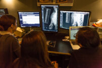
MRI also uses intravenous contrast agents to differentiate various soft tissues in the body. The MRI image you see on the right shows a tumor of the heart in a dog following administration of a contrast agent.
Your Pet and the Big Machines
Here are some tips for pet owners whose pets require a CT scan or MRI:
- Expect blood tests and possibly a chest x-ray to be done before the scan. Testing helps veterinarians determine safe anesthetic protocols for your pet.
- Unlike when you or I receive an MRI or CT scan, you should anticipate that anesthesia will be administered to your pet. You know how hard it is to get a clear photograph of your wiggly pet. We need them to be perfectly still for imaging so that we can obtain an accurate scan.
- Know that it may take up to 24 hours for the radiologist to issue a final report on the scan. Waiting is hard, but reviewing images takes time and should not be rushed.
