


Medical Machines: Ultrasound Machines
Medical Machines: Ultrasound Machines
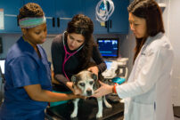
“Medical Machines” is a new series of blog posts highlighting the equipment AMC veterinarians use to provide state-of-the-art care to thousands of pets annually. These machines save countless lives, but pet families rarely ever have the opportunity to see them up close and personal. This series will give readers a glimpse into the equipment AMC veterinarians rely on every day. The previous blog post in this series highlighted infusion pumps.
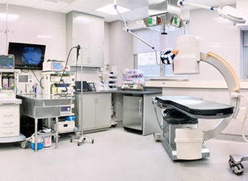
The machine for today is an ultrasound machine. I was shocked when I finished counting our ultrasound machines — AMC has ten different ones located on four different floors of the hospital.
An internal view without x-rays
Fifty years ago, if veterinarians wanted to see the inside of the body, we took an x-ray. To take an x-ray, we use a machine that emits radiation, creating an image on film. An x-ray reduces a three-dimensional dog or cat to a two-dimensional image. While x-rays are still used for the heart, lungs and bones, ultrasound is more commonly used to view the inside of the abdomen. The probe of the ultrasound machine bounces sound waves off the internal organs as the probe is moved about. The sound waves then create a gray scale image on the ultrasound screen. The veterinarian performing the ultrasound twists the probe which changes the orientation of the image and allows the various organs to be evaluated from side to side, top to bottom and front to back. By analyzing an organ in multiple directions, a clear picture of any structural abnormalities emerges.
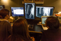
Diagnostic images = radiology
Back when veterinarians only had x-rays, the department in charge of x-rays was called radiology. Now that we have x-rays, CT scans and MRIs, the department has expanded and these imaging modalities reside in AMC’s Diagnostic Imaging Department. Since ultrasound is another method of imaging the body, this group of AMC doctors has two of our best ultrasound machines in the hospital. Our radiologists have special training to perform and interpret ultrasounds. One of their most important skills is using the ultrasound to collect samples for laboratory testing.
Peering into a heart
An x-ray of the chest will identify an enlarged heart or fluid in the lungs as the result of heart failure. To hone in on problems with heart values or a thickened heart muscle, a specialized ultrasound called an echocardiogram is required. AMC’s cardiologists have two such machines: one about the size of a small refrigerator and a portable one for cage side use.
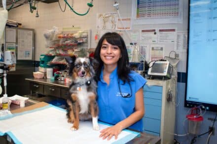
A quick look
The other six ultrasound machines are distributed on four different floors of the hospital, necessitated by AMC’s vertical space. All six of these machines have similar functions. They are quite small, about the size of a toaster and are strapped to a wheeled stand. ICU and ER doctors will wheel their machine to a critical patient to quickly determine if there is internal bleeding or an object stuck in the intestine. Oncology and Interventional Radiology use their mobile ultrasound machines to monitor the effect of treatment, as does Internal Medicine. We all use our ultrasound machines to facilitate collection of urine samples.
With all these essential functions, no wonder AMC needs a denary of ultrasound machines!