Canine Mast Cell Tumors: What You Need to Know
Canine Mast Cell Tumors: What You Need to Know
One of the most vexing problems for dog families is the skin lump. It could be a warty growth on the skin, a smooth moveable lump just under the skin or maybe a hard lump on the leg. Without a test, it’s impossible to know if it a just a benign fatty tumor or a malignant tumor. Since November is National Pet Cancer Awareness Month, today’s blog will focus on the most common malignant skin tumor of dogs, the canine mast cell tumor.
A what tumor?
If your veterinarian says your dog has breast cancer or lymphoma, you most likely have heard of those tumors before and have some idea where the tumor is. This is not typically the case with a mast cell tumor. Mast cell tumors are common in dogs, but rare in humans. Mast cells are the allergy cells responsible for puffy eyes and itching that comes with hay fever season. This explains why mast cell tumors sometimes puff up then magically get smaller on their own and why dogs often lick or scratch at mast cell tumors.
Which dogs are more likely to develop mast cell tumors?
Any dog can develop a mast cell tumor and the Cancer Institute at AMC cares for many mixed breed dogs with mast cell tumors, but certain breeds are at greater risk for developing mast cell tumors. One study identified these breeds as: Shar-Peis, American Staffordshire Terriers, Boxers, Labrador Retrievers, French Bulldogs and Golden Retrievers. There is no difference in the occurrence of this tumor between male and female dogs.
What does a mast cell tumor look like?
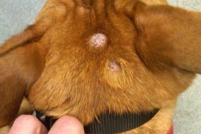
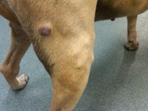
The photograph of a dog’s head above, shows two mast cell tumors. They are pretty typical ones: round, raised, hairless masses. If these had been larger, I would have expected them to be ulcerated and bleeding. The photograph of the hind leg of a different dog shows another typical mast cell tumor, but notice the entire leg is lumpy. The mast cells in this tumor are causing an allergic reaction and this dog has hives.
What kind of treatment is the best for mast cell tumors?
Veterinary oncologists use all available treatment options in the management of canine mast cell tumors. The most important treatment for mast cell tumors is surgery. In order to remove the entire tumor, the surgical recommendation is for there to be 3 cm margins removed around the tumor, so this procedure typically requires long incisions. If the biopsy indicates the tumor has not been completely removed or if the tumor cannot be removed at all, veterinary oncologists will likely recommend radiation therapy to control the tumor. If the biopsy indicates the mast cell tumor has a high likelihood of spreading, then chemotherapy is also recommended.
What are tumor grades 1, 2, 3?
When a mast cell tumor is removed, it’s sent to the pathologist for biopsy. The pathologist looks at the tissue and makes a variety of assessments. In the case of mast cell tumors, they assess how abnormal the cells look, how much the cell invades into normal tissue, how many cells are dividing and what the center or nucleus of the cell looks like. The pathologist then assigns a number: 1, 2, or 3. This is called a tumor grade. The more abnormal the cells are, the higher the tumor grade. In the oncology world, higher is bad and more treatment is required. In the case of mast cell tumors, high also indicates a less favorable prognosis. So if your pup has a skin lump, be sure to call it to your veterinarian’s attention, and remember: a test is the only way to know if the lump should be ignored or removed.
