AMC Loves Radiology and This is Why
AMC Loves Radiology and This is Why
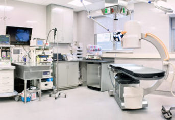
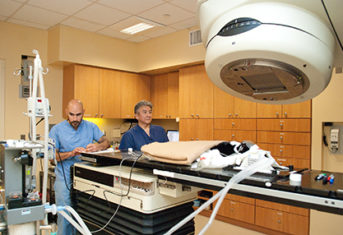
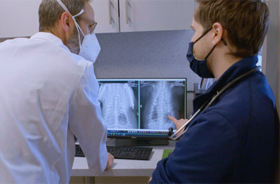
Although they’re out of view of many clients, the Diagnostic Imaging and Radiology Service is vital to the everyday functioning of the Schwarzman Animal Medical Center. Animals can’t tell us what’s wrong when they are sick or injured, so veterinarians must use a combination of expertise, experience, and technology to diagnose and treat their patients. AMC’s cadre of board-certified radiologists and radiology residents training for board certification support AMC’s veterinary specialists with a variety of imaging tools to help guide diagnoses and treatment plans, including x-rays, ultrasound, CT scans and MRI. On average, AMC veterinarians order 25 x-ray studies per day, with each study including at least 2 images.
Today’s blogpost will focus on some recent patients whose x-rays revealed the pet ate something they shouldn’t have and required urgent surgical removal by AMC surgeons to save their lives.
Magnamosis: A Potentially Fatal Attraction
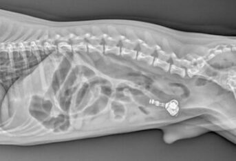
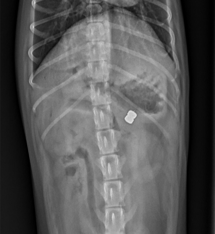
The first patient saved by an x-ray was a two-year-old terrier that almost didn’t make three. Trevor the terrier snacked on some small magnets. After he swallowed them, one stayed in the stomach and the other moved to the intestine. At some point, they were close enough together that the magnetic attraction pinched the stomach and segment of intestine to each other. In the x-ray, you can see the two stacked magnets clearly.
Trevor’s case is an example of magnamosis. Magnamosis is the use of special medical magnets to create an opening between two segments of intestine or two blood vessels. However, in Trevor’s case, the magnamosis was pathologic, causing perforation of both the stomach and intestine, peritonitis and intestinal obstruction. Fortunately, AMC surgeons were able to remove the magnets and repair the stomach and intestine. Other veterinarians have seen magnamosis cause serious illness in dogs. Never let pets play with or around magnets.
A Good Radiologist Needs Only a Kernel of Evidence
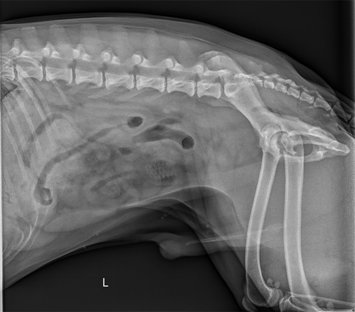
Saint is a known garbage eater. When he came to AMC’s ER with a complaint of vomiting for several days and a poor appetite, his first stop was the Radiology Service for an abdominal x-ray. Thanks to their skill, experience, and intuition, they spotted the cause without delay, which could have been invisible to a non-specialist. When a garbage-eating, vomiting dog shows up in the ER during the summer months, they know to hunt carefully for evidence of a corn cob foreign body. And there it was! Near the bottom of the abdominal x-ray, you can see a piece of corn cob with the kernels eaten off. The necessary surgery was successful, and Saint was quickly back to looking for a new snack from the trash can.
Foreign Body Obstruction is the Pits
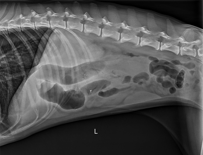
Kiara is normally a very energetic and hungry dog, but her family brought this 2-year-old Husky to AMC because she wouldn’t eat and was extremely lethargic. Because she had vomited a few times, the AMC ER staff ordered an x-ray. This x-ray is not as obvious as the magnet x-ray. The large black ovoid areas are gas filled intestinal loops, which clue veterinarians to look for an intestinal obstruction. AMC’s radiologists identified a hazy area in the center of the abdomen as a possible location of obstruction. Sure enough, emergency surgery confirmed an intestinal obstruction by a mango pit! Not surprisingly, the hazy area in the center of the abdomen is about the same size as a mango pit.
Has this blog piqued your interest in veterinary radiology? Explore some more patient x-rays here or see what happens when your dog eats your earbuds!
