


Celebrating World Kidney Day by Looking at Kidneys
Celebrating World Kidney Day by Looking at Kidneys
Usually I celebrate World Kidney Day by writing about kidneys. This year I am switching it up and am showing pictures of kidneys. In keeping with this year’s theme “Kidney Health for All,” I am going to show you how images from the Schwarzman Animal Medical Center’s Diagnostic Imaging Service help to ensure your pet’s kidneys are healthy using x-rays, ultrasound, CT scans and MRIs. AMC’s Diagnostic Imaging Service is a favorite in the hospital because we all rely on them to help us take care of your pets.
Radiology by Another Name
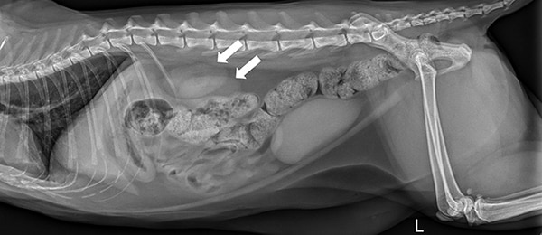
Some readers might be puzzled by the term diagnostic imaging. This is the more modern name for radiology. Yes, veterinarians still take x-rays, as you can see below. This is an abdominal x-ray of a cat taken from right to left. The kidneys are two oval, overlapping structures just behind the rib cage, identified by arrows.
Sonogram for Viewing Kidneys
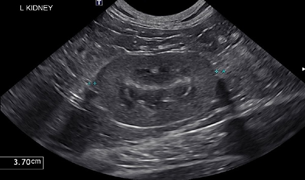
AMC veterinarians usually choose an ultrasound more often than x-rays to evaluate kidneys. Ultrasounds make images using sound waves. On an ultrasound, each kidney can be viewed separately, stones are easily visualized, and the internal structure of the kidney can be assessed. The measuring instruments built into the ultrasound machine measure this cat’s kidney at 3.7 cm (about 1.5 inches) in length and you can easily see the outer cortex of the kidney compared to the inner core called the medulla.
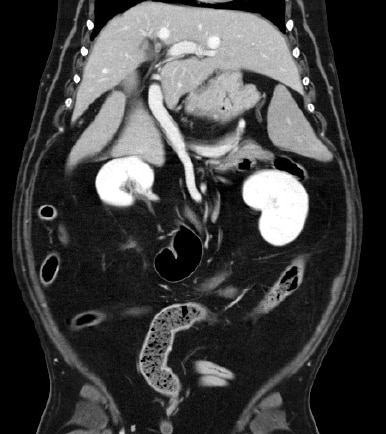
CT Scan Highlights Blood Vessels
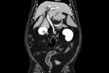
AMC veterinarians might use a CT scan to evaluate the blood supply of an organ. This CT scan of a dog’s abdomen to the right was performed using a special intravenous dye that makes the blood vessels bright white. Because the dye is excreted from the body by the kidneys, they show up as easily identifiable bright white structures as well.
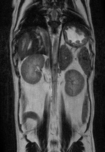
MRI Shows a Different Kidney Picture
The MRI of a dog’s abdomen to the left gives a completely different view of the kidneys. MRI or magnetic resonance imaging creates images with a magnetic field. X-rays and CT scans use radiation to create images. In this MRI view the kidneys appear as negative images and almost fade into the background. They retain their classic “kidney bean” shape seen in the other images in this blog.
I hope you enjoyed this glimpse of how veterinarians can examine your pets kidneys even though we can’t see them. Diagnostic images are just one way we work to keep the kidneys of your favorite fur person healthy.
About the Author
Related Posts
-
Diagnostic ImagingAugust 03, 2022

AMC Loves Radiology and This is Why
Learn More -
Internal MedicineMarch 09, 2022

CKD, BUN, USG, and AKI: How to Talk to Your Pet’s Veterinarian About Kidney Disease
Learn More -
Cats Internal MedicineJune 17, 2020

Chronic Kidney Disease in Cats and Subcutaneous Fluids
Learn More