Interventional Radiology & Endoscopy
About Interventional Radiology & Endoscopy at AMC
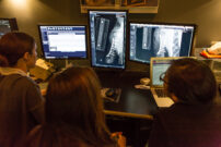
The Katharine and William Rayner Interventional Radiology and Endoscopy Suite is the first facility to implement a complete interventional service for veterinary patients. Interventional Radiology and Interventional Endoscopy are well-established technologies in human medicine, and our team has pioneered the use of these lifesaving procedures in companion animals.
Disorders treated interventionally at AMC include:
- Kidney, ureteral, and bladder stones
- Tracheal collapse
- Various tumors
- Canine incontinence
- Liver shunts
- Blood clots
- Narrowing of nasal passages
- Feeding tube placement
Veterinary Interventional Endoscopy
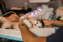
Interventional endoscopy (IE) involves the use of endoscopic equipment with other contemporary imaging modalities, such as fluoroscopy and/or ultrasound, to perform diagnostic and therapeutic procedures in virtually any part of the body accessed endoscopically (gastrointestinal, biliary, respiratory, urinary tract, etc.).
The combination of endoscopy and fluoroscopy allows veterinary technicians to access small orifices that would otherwise require more invasive surgical techniques. For example, endoscopic procedures are used to treat conditions such as:
- Dog and cat urinary tract infections
- Dog incontinence
- Kidney stones
- Respiratory tumors
- Nephrostomy tube placement
Veterinary Interventional Radiology
Interventional radiology (IR) involves the use of contemporary imaging modalities (CT scans) to gain access to different structures of the pet’s body for diagnostic and therapeutic reasons. Veterinary radiology refines similar procedures in human medicine to provide non-surgical care alternatives.
IR procedures have the potential to provide alternatives for our patients in whom conventional therapies are declined, not indicated, or associated with high risk of mortality. Pet radiology can be applied to any body system in patients of all sizes and is associated with:
- Better patient outcomes
- Minimal anesthesia time
- Reduced hospital stays
In addition, some techniques, such as chemoembolization of tumors or palliative stenting for malignant obstructions, offer treatment options for patients with various conditions that may not be amenable to standard therapies.
Canine and Feline Portosystemic Shunts (PSS)
Portosystemic shunts are abnormal connections between the portal venous and the systemic venous systems, leading to a variety of clinical signs affecting the neurologic, urogenital, and gastrointestinal systems. These shunts are diagnosed based on patient signalment and history, physical examination, biochemical testing, and diagnostic imaging studies. Patients are initially treated medically and then the best outcomes are provided through surgical or interventional therapies, which can provide a good prognosis.
VMD, DACVS
Co-Director of The Katharine and William Rayner Interventional Radiology and Endoscopy Service
Service Head of Interventional Radiology and Endoscopy
DVM, DACVIM (SAIM)
Co-Director of The Katharine and William Rayner Interventional Radiology and Endoscopy Service