Brachycephalic Airway Syndrome in Dogs

Background
The term brachycephalic comes from the Greek words brachy, meaning “short” and cephalic, meaning “head.” Brachycephalic dog breeds have flat faces with shortened muzzles. Unfortunately, the shortened muzzles and snouts often mean that the throat and breathing passages are also undersized or flattened. The term Brachycephalic Obstructive Airway Syndrome, or BOAS, refers to multiple anatomic abnormalities that can lead to breathing difficulties and other health problems for these dogs.
As many as six anatomic abnormalities make up BOAS. Not all dogs have all six abnormalities, but the more a dog has, the greater their clinical signs. The table below lists the medical names for the abnormalities followed by their definition.
| Anatomic Abnormality | Definition |
| Stenotic nares | Nose holes are too narrow or collapse inward during inhalation |
| Extended nasopharyngeal turbinates | Air filtering bones inside the nose extend into the back of the throat |
| Elongated soft palate | Roof of the mouth is too long |
| Laryngeal collapse | Voice box collapses, making air passage difficult |
| Hypoplastic trachea | Windpipe is too narrow for the dog’s size |
| Everted laryngeal saccules | Pouches inside the voice box turn inside out and block airflow |
All of these anatomic abnormalities lead to a decrease in air flow in and out of the lungs. The abnormalities associated with BOAS cause affected dogs to easily overheat because they cannot effectively cool themselves through panting. Stress, anesthesia, and exercise are also difficult for these dogs. Finally, dogs with BOAS often have lower blood oxygen levels as compared to non-brachycephalic breeds.
Risk Factors
All brachycephalic dog breeds are at risk for BOAS. Common brachycephalic breeds include:
- Boston terriers
- Boxers
- Bulldogs (English and French)
- Cavalier King Charles spaniels
- Mastiffs
- Pekingese
- Pugs
- Shih Tzus
Signs
Signs of BOAS include:
- Noisy breathing, snoring
- Dyspnea (difficulty breathing)
- Retching or gagging
- Exercise intolerance
- Blue tongue or gums (lack of oxygen)
- Collapse following activity, excitement, or high heat/humidity
Diagnosis
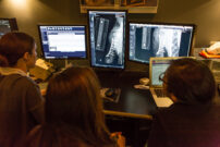
In order to definitively diagnose BOAS, the dog must be examined under anesthesia so the veterinarian can look for the anatomic abnormalities present in this syndrome. Stenotic nares (narrow nose) can be easily seen upon physical examination. The diagnosis of hypoplastic trachea is made using x-rays which also allows examination of your dog’s lower airways and lungs.
The University of Cambridge and The Kennel Club in the UK have also developed the Respiratory Function Grading Scheme (RFGS) to objectively measure the severity of BOAS in dogs and help veterinarians make a clinical diagnosis. The RFGS uses a scale from 0 to 3 to measure the severity of BOAS:
Grade 0: Clinically unaffected. Free of respiratory signs.
Grade I: Clinically unaffected. Dog has mild respiratory signs of BOAS but they do not affect exercise performance.
Grade II: Clinically affected. Dog has clinically relevant respiratory signs that require management.
Grade III: Clinically affected. Dog has severe respiratory signs and requires a thorough examination with treatment.
Treatment
Treatment of BOAS starts in the home. Since obesity will aggravate the issues already associated with BOAS, it is vital that your dog maintains an ideal weight and body condition score to make breathing easier. Additionally, pet owners should use a harness instead of a collar, which puts pressure on the voice box and windpipe.
Your veterinarian may prescribe anti-inflammatory medications, sedatives, and cough medications to help alleviate a flare-up of BOAS. There are also a variety of surgical interventions that may be needed to alleviate respiratory signs and provide long-term relief, especially if the signs are severe or are causing life-threatening obstructions. The table below describes the anatomic abnormality followed by the corrective surgery:
| Anatomic Abnormality | Corrective Surgery |
| Stenotic nares | Enlarge nostrils |
| Extended nasopharyngeal turbinates | Treatment is controversial and not widely available |
| Elongated soft palate | Shorten roof of mouth |
| Laryngeal collapse | Permanent tracheostomy, which creates an opening in the windpipe for breathing |
| Hypoplastic trachea | No surgical treatment available |
| Everted laryngeal saccules | Remove pouches blocking airflow |
Your pet will need to be closely monitored in the hospital following surgery, especially because inflammation or bleeding that occurs after a surgical procedure can obstruct the airway. Occasionally, a tube will be placed through an incision in the neck into the trachea to provide oxygen to the pet until the swelling subsides (tracheostomy).
In a recent research study from the Schwarzman Animal Medical Center, researchers determined a carbon dioxide laser technique that was as good as the bipolar vessel sealing device for surgical reduction of an elongated soft palate (shortening of the roof of the mouth). Both are preferable to traditional cutting and suturing because traditional surgical techniques result in more bleeding and swelling in this area.
Prevention
Unless you are a breeder who is able to exclude certain dogs from breeding with each other, BOAS cannot be prevented in dogs already born with these anatomic abnormalities. It is vital that you keep your pet at a healthy weight and body condition score. If you have a brachycephalic breed, make sure to keep them away from environments with high heat or humidity and be sure to watch them carefully as they engage in play or exercise. Should you notice your pet has difficulty breathing, take them to the veterinary ER right away.
Watch the Presentation
Dr. Daniel Spector, Service Head of Surgical Service 2 and the Surgery Residency Program Director at the Schwarzman Animal Medical Center, discusses the clinical signs of BAS, the various anatomic abnormalities that can cause breathing difficulties, and how it can be treated.
