February is Pet Dental Month: Part 2

February is Pet Dental Month: Part 2
The importance of dental care for dogs and cats.
Part 2 of a 3 part series by Stephen Riback, DVM
Like people, our pets are prone to dental disease. This month focuses on the importance of controlling and preventing dental disease in our cats and dogs. Untreated dental disease is associated with both infection and pain. Recent studies in people and dogs show that untreated infection in the mouth has also been linked to infections in other parts of their bodies.
Feline Odontoclastic Resorptive Lesions
The most common dental disorder in domestic cats is a decay of the teeth called Feline Odontoclastic Resorptive Lesions (FORLS). This is present in 50-75% of all cats presenting for dental procedures. We do not know what initiates the process, but we do know that a cell within the tooth called and odontoclast gets “turned on” and removes calcium, from within the tooth structure. These teeth then start to decay often from the inside out. This is different from “carious decay” or cavities in people that begins from outside surface of the tooth. When these lesions break through to the surface of the tooth, they become painful. The only approved treatment for FORLS is extraction of the affected teeth. Many cats are living with FORLS and do not show overt signs. Some cats may have difficulty eating dry food or avoid using teeth with FORLS present and some cats may have jaw “chattering” while chewing.
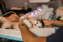
The diagnosis and treatment of FORLS involves general anesthesia. Animals should be prescreened to ensure they are good anesthetic candidates before undergoing a dental procedure. This often involves a thorough physical exam, blood tests and sometimes x-rays of the heart and lungs, or an echocardiograms of the heart. Once a patient is safely anesthetized, the teeth should be cleaned to evaluate for dental disease. Probing around every surface of every tooth in a cat will screen for FORLS. In the early stages, a FORL may look like a small hole within the tooth. As the FORLS progress, more destruction of the tooth may be present. Many times, gum tissue will grow over the exposed FORL giving the appearance of the gums growing onto the tooth surface. “Intra-oral” or dental radiographs can also be taken to help diagnose FORLS. Many practices now have “digital” or computerized dental X-Rays that do not need dental films and require much less radiation than the old X-Rays. The combination of dental probing and X-Rays can help determine the best treatment for an individual patient. Many cat owners report that after successfully extracting teeth with FORLS, their cats seem happier and more active. Cats seem to thrive better with extracted teeth, versus painful teeth.
Some FORLS seem to be associated with inflammation at the gum line. Many FORLS will develop independent of gingival inflammation. Keeping gingivitis under control may help prevent some FORLS, but many are not preventable. Cats who develop FORLS in some teeth will usually develop new FORLS in other teeth in the future, so annual oral examinations are recommended to identify new lesions as they develop.
Readers Poll:
[polldaddy poll=1375345]
Check back for results!
—————————————-
About Stephen Riback, DVM
Dr. Riback received his veterinary degree from the New York State College of Veterinary Medicine at Cornell in 1985. He was a general practitioner from 1985 until 1999 and owned the Oakdale Veterinary Hospital from 1989 until 1999. Dr. Riback has worked at the AMC since 1999, first in the Community Medicine dept. and then from 2003 in the Dentistry dept. where he studied dentistry under the mentorship of Dr. Dan Carmichael, who is the only board certified veterinary dentist in New York City.
The department of dentistry is the only full service veterinary dental practice in New York City and operates Monday through Friday at the AMC. Dr. Carmichael works on Mondays and Dr. Riback is in Tuesday through Friday. Dental procedures and oral surgeries are performed Monday through Friday.
