Medical Glue to the Rescue: Correcting Abnormal Blood Vessels
Medical Glue to the Rescue: Correcting Abnormal Blood Vessels
An important part of AMC’s mission is creating new medical knowledge. Our veterinarians advance the care and treatment of companion animals by conducting innovative clinical research and publishing their results in veterinary journals. This blogpost highlights research by AMC’s Interventional Radiology Service and Surgery Department.
In prior blogposts about research publications from AMC clinicians, I highlighted one of our most common surgical procedures: the repair of torn cruciate ligaments. Another research-focused blogpost highlighted AMC’s Emergency Room Fast Track service and how it has inspired Fast Track services in other veterinary hospitals throughout the country. Today’s blogpost reports on an uncommon condition, arteriovenous anomalies, with a creative correction.
Normal Arteries and Veins
To understand an arteriovenous anomaly, you need to understand the normal blood vessel anatomy in dogs and cats. Arteries are under high pressure as they speed blood away from the heart, and veins are under lower pressure as they return blood to the heart. These vessels typically lie side by side throughout the body, but they do not connect. An arteriovenous anomaly occurs when the artery and vein develop an abnormal connection, resulting in aberrant blood flow.
Arteriovenous Anomaly of the Leg
Arteriovenous anomalies are rare and present a diagnostic challenge. An animal with an arteriovenous anomaly may have a swollen leg covered with distended, disorganized and discolored blood vessels under the skin. The abnormal connection between the high-pressure artery and low-pressure vein shunts the blood from the artery to the vein, bypassing the “downstream” blood vessels towards the end of the limb. Because normal blood flow is disrupted, the leg swells.
A Picture of Arteriovenous Anomaly is Worth a Thousand Words
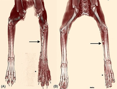
The photographs accompanying this blogpost are from the publication entitled, “Dominant outflow vein occlusion in the management of naturally occurring peripheral arteriovenous anomalies in cats and dogs.”
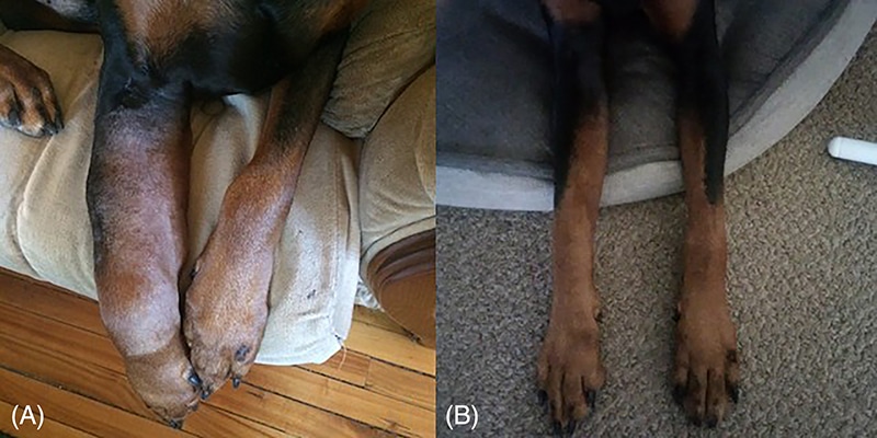
The left panel of each illustration shows one of the dog patients in this study before correction of the arteriovenous anomaly. You can see the distended blood vessels and generalized swelling typical of an arteriovenous anomaly. The panel on the right shows the same dog after the arteriovenous anomaly has been corrected: a magical transformation. How did this happen? The title of the article says it all, except in a difficult-to-understand way for non-specialists. Here’s the backstory. AMC’s Interventional Radiology team used CT angiography to identify the major vein leading out of the arteriovenous anomaly and used a minimally-invasive surgical procedure to plug it up with a suture or medical glue. Blocking the major outflow vein rerouted the blood through a more normal pathway. And voilà, the swelling was gone!
Congratulations to AMC veterinarians Drs. Weisse and Schwartz and AMC alum Dr. Hyndman on their clinical and research successes.
